Safe, painless cardiac imaging using ultrasound — evaluating your heart's structure, valves, and function in real time, with clear reporting you can understand.
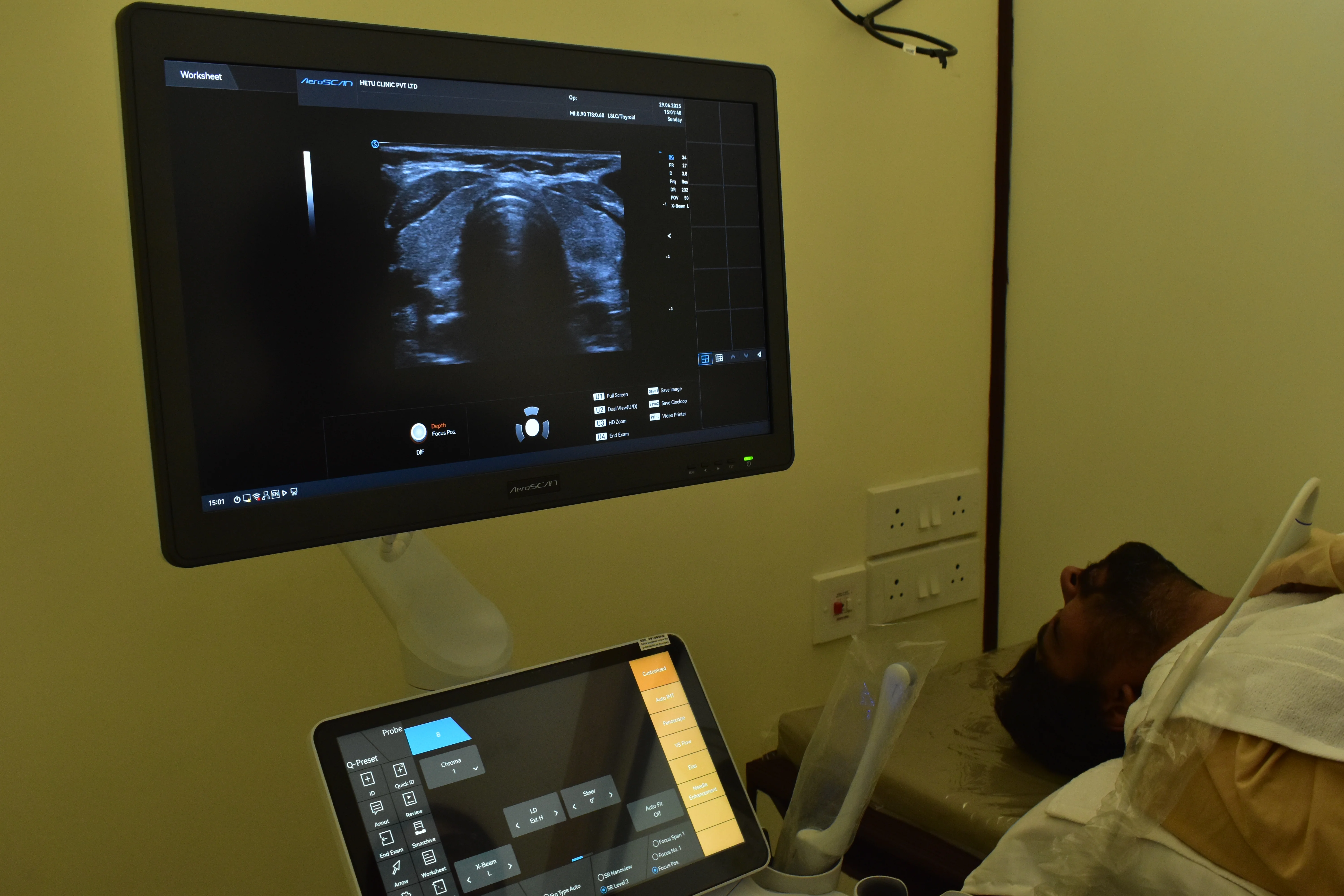
2D Echocardiography — commonly referred to as 2D Echo — is a safe, non-invasive imaging test used to evaluate the structure and function of the heart. It uses ultrasound waves to create real-time images of the heart's chambers, valves, walls, and blood flow patterns, providing critical insights into cardiac health.
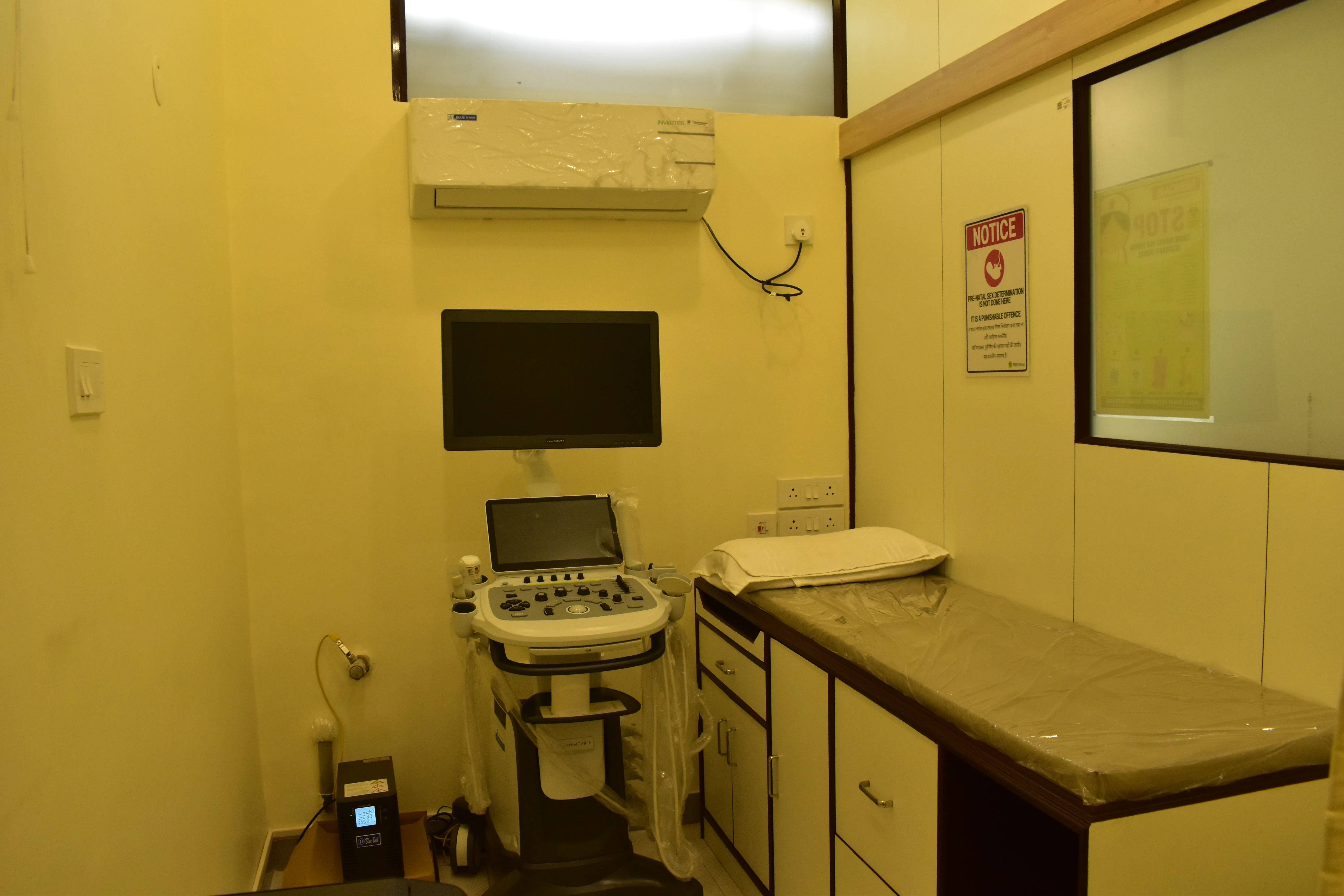
At Hetu Clinic, our 2D Echo studies are conducted by experienced professionals using high-resolution equipment. The test is painless, does not involve radiation, and usually takes 20–30 minutes.
Your physician may refer you for a 2D Echo in a range of cardiac and non-cardiac conditions.
Evaluation of unexplained chest pain, breathlessness, or palpitations to identify underlying cardiac causes.
Diagnosis and monitoring of valve regurgitation, stenosis, and other valvular abnormalities over time.
Assessment of heart function in patients with high blood pressure or diabetes — detecting early cardiac changes.
Assessing residual cardiac function, wall motion abnormalities, and complications following a myocardial infarction.
Screening and monitoring of structural defects present from birth — in children and adults alike.
For patients over 40 or those with a family history of heart disease seeking a baseline cardiac assessment.
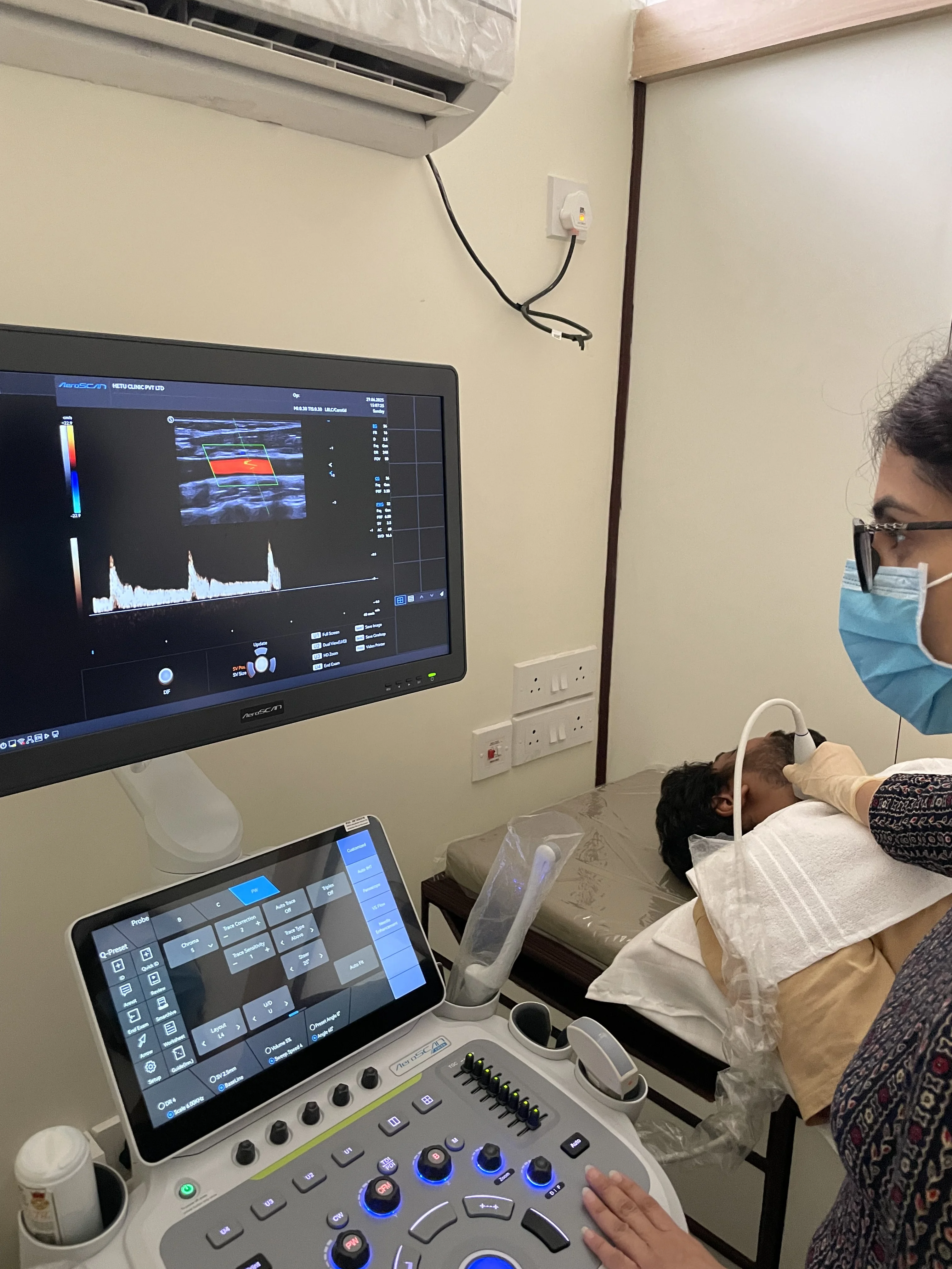
We offer both plain 2D Echo and Colour Doppler studies for comprehensive cardiac assessment.
Visualises heart structures and motion in real time. Evaluates chambers, walls, and valves — the foundation of cardiac imaging.
Adds blood flow mapping to assess the direction and speed of flow — essential for diagnosing valve regurgitation, stenosis, and congenital anomalies.
Appointments are available on prior booking. Walk-ins may be accommodated based on radiologist availability. Reports are explained clearly.